| name |
Amanita lavendula |
| author |
Tulloss, K. W. Hughes & Rodríg.-Cayc. in Tulloss
et al. 2015.
Amanitaceae 1(2): 2. |
| name status |
nomen acceptum |
| english name |
"Coker's Lavender Staining Amanita" |
| synonyms |
≡ Amanita citrina f. lavendula(Coker) Veselý. 1933. Ann. Mycol. 31(4): 239.
≡ Amanita mappa var. lavendula Coker. 1917. J. Elisha Mitchell Scient. Soc. 33(1/2): 39, pl. 22-23, 64.
≡Amanita citrina var. lavendula (Coker) Sartory & L. Maire. 1922. Compend. Hymenomyc.—Amanita ??: 25.
≡ Amanita porphyria var. lavendula (Coker) L. Krieg. 1927. Mycologia 19: 309.
≡ Amanita brunnescens f. lavendula (Coker) E.-J. Gilbert nom. inval 1941. Iconogr. Myco. (Milan) 27 suppl. 1 (1-3): 336. [Not accepted by author in original publication. ICBN §34.1(a)]
The editors of this site owe a great debt to Dr. Cornelis Bas
whose famous cigar box files of Amanita nomenclatural information
gathered over three or more decades were made available to RET for computerization
and make up the lion's share of the nomenclatural information presented on this site.
|
| MycoBank nos. |
508879, 166594, 508880, 171834, 807618 |
| GenBank nos. |
Due to delays in data processing at GenBank, some accession numbers may lead to unreleased (pending) pages.
These pages will eventually be made live, so try again later.
|
| lectotypes |
NCU |
| lectotypifications |
Tulloss et al. 2015. Amanitaceae 1(2): 2.
Rejected lectotypification of Neville and Poumarat
(2004: 818)
rejected by Tulloss (2005a.
Mycotaxon 92: 480 [footnote]). |
| intro |
The following text may make multiple use of each data field.
The field may contain magenta text presenting data from a type study
and/or revision of other original material cited in the protolog of the present taxon.
Macroscopic descriptions in magenta are a combination of data from the protolog and
additional observations made on the exiccata during revision of the cited original
material.
The same field may also contain black text, which is data from a revision of the present
taxon (including non-type material and/or material not cited in the protolog).
Paragraphs of black text will be labeled if further subdivision of
this text is appropriate.
Olive text indicates a specimen that has not been thoroughly examined (for example, for microscopic details) and marks other places in the text where data is missing or uncertain.
The following morphologcial material not cited as
the work of another researcher is based on original
research of R. E. Tulloss. Phylogenetic analyses
are based on
continued original research in the laboratories of
Drs. K. W. Hughes (Univ. of Tenn., Knoxville) Dr. L. V.
Kudzma (Annandale, New Jersey).
All parts of the fruiting body are liable to become
lavender or a similar purplish shade when ambient
temperature is below ca. 1° C for several
hours. This character is shared with the two
other known macromorphologically similar taxa in
eastern North America—A. cornelihybrida and
A.
americitrina. |
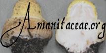
| pileus |
protolog: "...from 3.5 to 8
cm in diameter, flat or slightly depressed in
center (sometimes slightly gibbous in center) a
light but distinct primrose yellow (not the dull
egg yellow shades of A.
russuoloides), often with stains of light
brown, lavender, purple lavender, or a combination
of these; somewhat viscid when damp, shining when
dry"; context "...nearly white, sometimes
quickly turning to shades of lavender when cut,
quite thin, only 2 mm thick at center of gills...";
margin "disctinctly striate when mature, or
the striae may be scarcely visible until the margin
begins to dry," not appendiculate; universal
veil as occasional flat, irregular, lavender or
pink-lavender patches."
RET: 32 - 115 mm wide, 4A3 at margin and 3B3 over disc or
very pale citrine-yellow except brownish over disc or
mostly ivory and tinged with tan over disc,
becoming paler (except over disc) with age,
planoconvex with slight umbo to planar, sometimes
deeply rimose, tacky to wasy, shiny; context
3 - 5 mm thick, white to grayish white, sometimes
with watersoaked line above lamellae and above join
with stipe context, thinning evenly to margin;
margin not striate, not appendiculate;
universal veil absent or in scattered flat
patches, 5B2 to grayish. |
| lamellae |
protolog: "...pure white, free
but close to the stem and connected by a line which
runs down a little way [on the apical region of the
stipe], deepest near their middle, where they were
from 3.5 to 7 mm deep [i.e., broad], deep according
to the size of the plant [sic]..., none forked.";
lamellulae "..many...."
RET: free, without decurrent line at stipe apex,
close to crowded, sordid white to cream to ca. 3A2 in
mass, white to pale watersoaked cream and sometimes
becoming slightly sordid in age in side view, 2.5 -
6.5 mm wide, broadest ca. midlength;
lamellulae subtruncate to truncated
(shortest) or subattentuate to attenuate
(longest). |
| stipe |
protolog: ?...up to 10 cm
long and from 6 to 10 mm thick in center, smooth
and somewhat silky-shining, faint primrose-yellow
[appearing to be of substance continuous with
partial veil, which breaks up and remains
concolorous with partial veil, but fades] above and
nearly white below the veil, but often with cream,
brown, or lavender tints, and brown where bruised";
context "solid but sometimes nearly hollow
from the separation of the looser central fibers,
no distinct central cylinder"; bulb “"large
and abrupt, but variable, sometimes 2.8 cm in
diameter, soft and spongy generally with an
abruptly truncated top, which may be quite smooth
or show slight marginal projections representing
the volva"..."the surface is a distinct lavender
color, sometimes pinkish or brownish lavender or
rarely nearly white (as in No. 1399, but even in
this collection the volva patches on the cap were
lavender), internally it is white"; partial
veil "primrose yellow, thin, delicate, but not
flocculent or friable, the lower side often showing
the fibers by which it was attached to the
stem...it remains attached to the stem from 2 to
3.5 cm below the cap, generally collapsing tightly
against the stem, and so delicate at times that it
is scarcely noticeable on the mature plant [sic]
except where its free edge marks out a colored line
against the stem. At other times the veil remains
expanded for some time as a perfect skirt, and is
quite perfect in the mature plant [sic]";
universal veil "there is no volval
cup."
RET: 80 - 163 × 6 - 17 mm, white below partial veil,
pale citrine-yellow above, becoming grayish with age
or handling, rusty brown in old wounds, narrowing
upward (sometimes only slightly) or cylindric,
slightly to markedly flaring at apex, pruinose and
similar to upper surface of partial veil above
latter, below satiny and striatulate at first,
sometimes becoming subshaggy-fibrillose at least in
lower third of stipe; bulb 10 - 23 × 13 - 24
mm, subglobose, sometimes with rusty stains on
exterior; context stuffed or solid, off-white,
with rusty marks in old wounds, with central
cylinder 4 - 8.5 mm wide and sometimes becoming
watersoaked, insect tunnels not observed;
partial veil membranous, persistent,
skirt-like, superior, pale yellow, becoming
grayish from edge inward with age, faintly striate
above (at least near edge), bottom surface
concolorous with upper surface and bearing
subrectangular fragments of graying universal veil
limbus internus around the edge; universal
veil as brief and irregular circumcissile limb
separated from stipe by sinus ca. 2± mm
wide, white at least at first, with color change
similar to that on pileus. |
| odor/taste |
protolog: Odor "...when
freshly cut like raw green peanuts."
RET: Odor like freshly dug potatoes.
Taste not recorded. |
macrochemical
tests |
Spot test for tyrosinase (0.1% paracresol) - negative throughout basidiome for both youngest and oldest specimens tested. Spot test for tyrosinase (L-tyrosine) - negative throughout basidiome for specimen just prior to maturity. Test vouchers: Tulloss 11-30-85-E, Tulloss 7-18-96-F, 9-26-97-D. |
| lamella trama |
bilateral, divergent; ??. |
| subhymenium |
pseudoparenchymatous (cellular); ??. |
| basidia |
?? × ?? μm, dominantly 4- and infrequently 1- or 2-sterigmate, with sterigmata ?? × ?? μm; clamps not observed. |
| stipe context |
longitudinally acrophysalidic: ??. |
| lamella edge tissue |
sterile; ??. |
| basidiospores |
composite data from original material, RET:
[40/2/2] (5.8-) 6.0 - 7.5
(-8.1) × (5.1-) 5.2 - 6.5 (-7.0) μm, (L=
6.4 - 6.8 μm; L' = ?? μm; W = 5.8 -
6.0 μm; W' = ?? μm; Q = (1.03-) 1.05 - 1.22
(-1.25); Q = 1.11 - 1.13; Q' = ??),
??, ??, thin-walled, smooth, amyloid, subglobose to
broadly ellipsoid, adaxially flattened;
apiculus sublateral, cylindric, varying in
width; contents granular; white in
deposit.
composite data from all collections revised by
RET & CRC: [140/7/6] (5.8-) 6.0 - 8.0 (-9.7)
× (4.9-)
5.3 - 7.0 (-8.1) μm, (L = 6.5 - 7.2 μm;
L' = 6.9 μm; W = 5.8 - 6.4 μm;
W' = 6.1 μm; Q = (1.02-) 1.05 - 1.29
(-1.49); Q = 1.10 - 1.18; Q' = 1.13),
hyaline, colorless, thin-walled, smooth, amyloid,
subglobose to broadly ellipsoid, occasionally
globose, infrequently ellipsoid, predominantly
adaxially flattened; apiculus sublaterial,
cylindric; contents predominantly guttulate,
occasionally granular; white in deposit. |
| ecology |
Subgregarious to solitary. La Vega, Dominican
Republic: Under Pinus occidentalis.
Hidalgo, México: In friable dark soil of
mesophytic Quercus-Pinus forest
(at least sometimes Pinus patula).
Connecticut: In loam and plant litter of deciduous
forest with Quercus alba, Q. rubra,
Betula sp., Fagus grandifolia,
etc.
Missouri: A pair. At 262 m elev.
New Jersey: Clustered to gregarious. At 35 m
elev. In deep sandy soil of
Pinus rigida-Quercus barrens or in
similar soil under Pinus.
South Carolina: In loamy soil of
Pinus-Quercus forest. |
| material examined |
CANADA:
ONTARIO—Norfolk Co., Port Dover
[42.7863° N/ 80.198° W, 182 m], 1.ix.2013 Eva
Skific s.n.
[mushroomobserver.org
#144353]
(RET 566-3, nrITS seq'd.); Walsh
[42.7616° N/ 80.394° W, 223 m], 28.ix.2013 E.
Skific s.n. [mushroomobserver.org
#146683]
(RET 575-6, nrITS seq'd.), 3.viii.2014 Eva
Skific s.n. [mushroomobserver
#172318]
(RET 639-7, nrITS & nrLSU seq'd.).
DOMINICAN REPUBLIC:
LA VEGA—N of Buena Vista, 4-5 km N
of Jarabacoa, along Nat. Rte. 28, 10.xi.2011 C.
Angelini ANGE95 (RET 693-2,
nrITS-LSU seq'd.).
MÉXICO: HIDALGO—Mpio.
Molango - Laguna Atezca [20°47’32” N/ 98°44’53” W,
1260 m], 18.vii.1996 Dr. Arturo Estrada Torres &
Lucía Varela Fregoso s.n. [Tulloss 7-18-96-H] (RET
253-2, nrITS seq'd.). Mpio. unkn. - ca.
northern jct. of Méx. Rte. 105 deviat. for
Zacualtipan & main Rte. 105 [20°40’28” N/
98°40’29” W], 18.vii.1996 Adriana Montoya Equivel,
A. Estrada Torres, L. Varela Fregoso & R. E. Tulloss
[Tulloss 7-18-96-E] (RET 252-7, nrITS seq'd.),
[Tulloss 7-18-96-F] (RET 252-10, nrITS seq'd.).
U.S.A.:
CONNECTICUT—Middlesex Co. - East Haddam,
Devil's Hopyard St. Pk. [41°28’57” N/ 72°20’30” W],
21.ix.1996 R. E. Tulloss 9-21-96-G (RET 250-3,
nrITS seq'd.).
MISSOURI—Ste. Genevieve Co. - W of Ste.
Genevieve, Hawn St. Pk. [37.8337° N/ 90.2416° W,
262 m], 30.x.2011 Patrick Harvey s.n.
[mushroomobserver.org #81042]
(RET 495-8, nrITS seq'd.), 14.x.2012 P. Harvey s.n.
[mushroomoberver.org #113456]
(RET 554-9, nrITS & nrLSU seq'd.),
[mushroomoberver.org #113460]
(RET 554-7, nrLSU seq'd.),
[mushroomoberver.org #113461]
(RET 554-8, nrLSU seq'd).
NEW JERSEY—Burlington Co. - Brendan T.
Byrne [=Lebanon] St. For., Pakim
Pond [39°52’49” N/ 74°32’02” W, 35 m], 9.x.1994
NJMA foray participant s.n. [Tulloss 10-9-94-F]
(RET 133-4, nrITS seq'd.), 26.x.1997 NJMA foray
participant s.n. [Tulloss 10-26-97-D] (RET 271-6,
nrITS seq'd.), 26.x.2008 Nina Burghardt s.n. (RET
447-4, nrITS seq'd.). Ocean Co. - ca.
Barnegat, Cedar Crk. or Oyster Crk Picnic Area,
Garden St. Pkwy., 9.xi.1989 Wm. Bakaitis 89-13 (RET
093-4, nrITS seq'd.); Waretown [ca. 39°47’12”N /
74°11’50” W], 5.x.1993 Cornelius Hogenbirk 10
(RET 111-4, nrITS seq'd.), 10.x.1993 C. Hogenbirk
12 (RET 111-6, nrITS seq'd.).
NEW YORK—Warren Co. - Warrensburg, Pack
Forest, 12.ix.1992 Wm. Bakaitis s.n. (RET 071-1,
nrITS seq'd.).
NORTH CAROLINA—McDowell Co. - GSMNP,
Cades Cove, Forge Crk. Rd., 27.ix.2006 D. J. Lodge
& E. Lickey s.n. [Tulloss 9-27-06-I]
(RET 396-8, nrITS seq'd.); ca. Little Switzerland,
Wildacres Retreat [35.8246° N/ 82.1065° W, 971 m],
30.ix.2006 Glenda O’Neal s.n. [Tulloss 9-30-06-C]
(RET 396-2, nrITS seq'd.).
Orange Co. - Chapel Hill,
Battle's Branch, short distance
above first bridge, woods,
27.x.1914 H. R. Totten s.n. [W. C. Coker 1432]
(lectotype, NCU, photo included in
protolog, spore print, unambiguous mention of
lavender in annotation); Chapel Hill, Howell's
Branch, and along branch from Strowd's, 18.x.1912
H. R. Totten 590 (NCU, photo included in protolog,
spore print, unambiguous mention of lavender in
annotation).
SOUTH CAROLINA—Anderson Co. - Hartwell
Lk., 27.xii.1982 Mary A. King & R. E. Tulloss
12-27-82-D (RET 399-4, nrITS seq'd.).
Oconee Co. - Seneca [34°46'09" N/ 82°57'55" W, 263
m], 30.xi.1985 R. E. Tulloss 11-30-85-E (RET 132-3,
nrITS seq'd.).
TENNESSEE—Blount Co. - GSMNP, Cades
Cove, Forge Crk. Rd., 27.ix.2006 D. J. Lodge &
E. Lickey s.n. [Tulloss 9-27-06-I] (RET 396-8,
nrITS seq'd.); GSMNP, Rabbit Crk. Tr., 2.ix.2006
unkn. coll. s.n. [TFB 12917] (TENN 61382, nrITS
seq'd.);. |
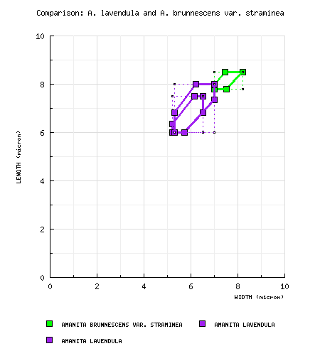
| discussion |
A comparison of sporographs based on the original
material of the present taxon that are candidates
for designation as its lectotype and
Amanita brunnescens var.
straminea, a clearly citrinoid taxon,
are found in the following diagram:
Note regarding lectotypification: Thirteen of 17
syntypes for A. mappa var. lavendula
were located by RET in NCU. These were
examined in order to support selection of a
lectotype from among them. In the process of
selecting a lectotype, RET first eliminated
material for which the annotation of fresh
material does not
unambiguously state that lavender was present on a
basidiome in a given collection. In addition,
a syntype was eliminated from consideration if it
is missing some significant element such as
universal veil on the bulb or the partial
veil. Existence of a photograph of the fresh
material and, especially, publication of such a
photograph of a given collection in the protolog
was viewed as increasing the value of the
collection, all other elements being equal.
When it became clear that the two spore size ranges
cited by Coker were associated with different spore
shapes and that the size range provided by Coker in
his basic description (rather than in a comment) was
associated with the best candidate for lectotype,
this was the final deciding factor in choosing a
collection to propose as lectotype (Tulloss et al
2015).
Note regarding molecular results (1.vi.2015 per
Hughes and Tulloss, personal communication): Recent
molecular studies indicate that collections
previously determined as A citrina in the
sense of eastern U.S. authors or A. citrina
var. lavendula can be divided into
three distinct taxa (segregated by nrITS
and nrLSU sequences).
All three taxa have been observed
to turn lavender upon exposure to near freezing or
freezing temperatures.
The collections
that have been segregated by sequencing are grouped
on this site on the present page and under the
temporary codes A.
cornelihybrida and A americitrina.
Notice that more than one of these
taxa can be found at a single location (e.g., Pakim
Pond in the New Jersey pine-oak barrens) and that
all three can be found in the sandy
soil of the New Jersey pine-oak barrens in the
Atlantic Coastal Plain.
The taxon here called "A. cornelihybrida" is
particularly curious because the samples of this
taxon instead of sharing a relatively invariant
nrITS sequence as in the other two cases, is
apparently an interbreeding cluster of
infraspecific hybrids (Hughes et al., 2013).
Hughes et al. hypothesize that the
ancestors of the cluster may have begun to diverge
genetically while in isolated glacial refugia;
however, this divergence did not lead to the point
of reproductive isolation. When the forests
and their symbionts moved outward from the refugia
at the end of the period(s) of glaciation, the
formerly isolated, lavender staining groups could
meet and mate—producing the infraspecific hybrid
cluster. At the present time, there is no
indication that the original genomes of the
isolated ancestors can be recovered from samples of
cornelihybrida; on the other hand the formerly
isolated genomes have not had the time to become
homogenized within cornelihybrida. Hughes et
al. have regularly extracted multiple distinct
sequences of nrITS (for example) from a single
basidiome of A. cornelihybrida.
Note: The illustration of the collection from North
Carolina (Tulloss 9-30-06-C) indicates that
a fruiting
body with rather strong yellow on the cap (as
opposed to the pallid cap described by Coker) can
have strong lavender staining and be assignable to
A. lavendula.
|
| citations |
Those working on this species acknowledge the support
of the University of Tennessee, Knoxville, and
National Science Foundation grant DEB 1144974.—R. E.
Tulloss and K. W. Hughes.
Remaining unsorted collections. Attention required:
non-type material:
U.S.A.:
MISSISSIPPI—Harrison
Co. - ca. Saucier, Choctaw Crk. Woods, 2.xii.1989
Anna Pleasanton s.n. [Tulloss 12-2-89-I]
(RET 145-10).
NEW JERSEY—Cape May
Co. - Belleplain St. For., Jakes Landing Rd.
[39.1892° N/ 74.8531°, 3 m], 21.x.2006 R. E. Tulloss
10-21-06-A (RET 398-1), -K (RET 397-9).
Ocean Co. - Waretown
[ca. 39.7867° N/ 74.1972° W, 3 m],
1983 Cornelius Hogenbirk 83-1
(RET 132-8).
NEW YORK—Suffolk
Co. (Long Isl.) - East Hampton Town, Napeague,
Cranberry Hole Rd., 17.x.1992 Lance T. Biechele
s.n. (RET 073-8).
SOUTH CAROLINA—Abbeville Co. - Sumter Nat. For.,
29.xii.1982 ?? 12-29-82-? (RET ??).
Anderson Co. - Hartwell Lake [34.3559º N/ 82.8217º
W, 161 m], 27.xii.1982 R. E. Tulloss
12-27-82-C (RET ??),
-E
(RET 399-5), -F (RET 399-10), -G (RET 399-9),
-H (RET 399-3).
Oconee Co. - Seneca [34°46'09" N/ 82°57'55" W,
263 m], 11-28-85-E (RET 132-9), 11-30-85-C (RET
131-2).
|
| editors |
RET |
|


, Chapel Hill, Orange Co.,
North Carolina, USA.")
, Chapel Hill,
Orange Co., North Carolina,
USA.")


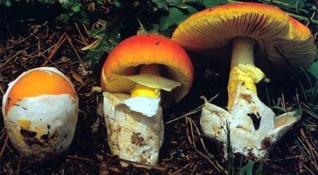
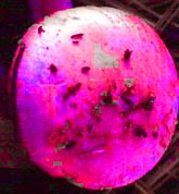 We continued the experiment with additional
photographs of other photographs of "A.
lavendula group" fruiting bodies
taken in December at the same South Carolina
site. The same phenomena was
observed—significant areas of the fruiting body
(especially those that had seemed lavender to the
naked eye) became strongly magenta. One of
the photos from the site shows extensive lavender
coloration over most of the cap (first row, right,
at the top of this page). In this case,
nearly the entire pileus became magenta as a result
of the maximum saturation experiment ("hypermagenta
test") (left). To date, the hypermagenta test
has been applied with negative results only to
photographs of citrina-like material
from Europe (France, Norway
and the United Kingdom) and to specimens of the
current taxon on which no lavender could be
detected by the eye. We are very interested
in testing photographs (for which there are dried
voucher specimens) from parts of Europe other than
those cited above.
We continued the experiment with additional
photographs of other photographs of "A.
lavendula group" fruiting bodies
taken in December at the same South Carolina
site. The same phenomena was
observed—significant areas of the fruiting body
(especially those that had seemed lavender to the
naked eye) became strongly magenta. One of
the photos from the site shows extensive lavender
coloration over most of the cap (first row, right,
at the top of this page). In this case,
nearly the entire pileus became magenta as a result
of the maximum saturation experiment ("hypermagenta
test") (left). To date, the hypermagenta test
has been applied with negative results only to
photographs of citrina-like material
from Europe (France, Norway
and the United Kingdom) and to specimens of the
current taxon on which no lavender could be
detected by the eye. We are very interested
in testing photographs (for which there are dried
voucher specimens) from parts of Europe other than
those cited above.

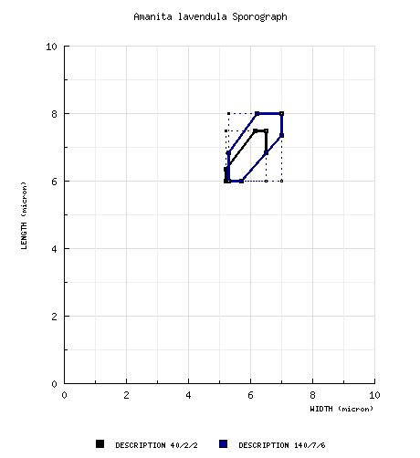
 Spore data sets and their composite